Etsuo Chihara1, Jin Ye Yeo2
1Sensho-kai Eye Institute, Kyoto, Japan; Shimane University, Izumo, Shimane, Japan; 2AES Editorial Office, AME Publishing Company
Correspondence to: Jin Ye Yeo. AES Editorial Office, AME Publishing Company. Email: aes@amegroups.com.
This interview can be cited as: Chihara E, Yeo JY. Meeting the Editorial Board Member of AES: Prof. Etsuo Chihara. Ann Eye Sci. 2025. Available from: https://aes.amegroups.org/post/view/meeting-the-editorial-board-member-of-aes-prof-etsuo-chihara.
Expert introduction
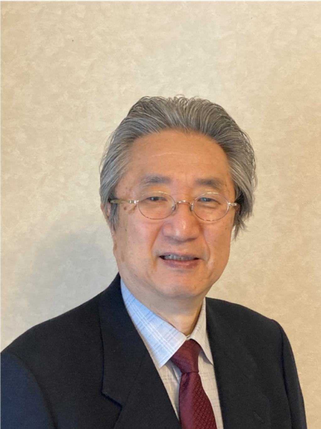
Prof. Etsuo Chihara (Figure 1) is a distinguished Japanese ophthalmologist. He graduated from Kyoto University and completed his residency in Ophthalmology at Kobe Central Municipal Hospital in Hyogo and Kurashiki Central Hospital in Okayama. His academic career commenced in 1977 at Kyoto University, where he earned his PhD in 1982.
He has authored over 100 peer-reviewed papers in English and more than 30 book chapters. He has been an invited speaker at over 100 national and international conferences and has served as the editor of 10 ophthalmology books. Notably, he presented the highest-class glaucoma lecture in Japan, the Suda Memorial Lecture, in 2012 and was elected an Emeritus Member of the Japanese Glaucoma Society in 2015. Prof. Chihara has also received numerous scientific awards.

Figure 1 Prof. Etsuo Chihara
Interview
AES: What motivated you to pursue ophthalmology and subsequently specialize in glaucoma surgery and imaging?
Prof. Chihara: I have had moderate deafness caused by otitis media since I was 10 years old, and I have long suffered from this issue. Therefore, I understand how crucial it is to receive information from the outside world.
Humans have five senses: sight, hearing, taste, touch, and smell. We receive 80 % of our information through vision, so maintaining good visual acuity is vitally important for a fulfilling life. This is the reason why I am eager to contribute to improving the visual function of patients.
AES: Could you provide an overview of the current publications in glaucoma surgery? Were there any articles or recent advancements that impressed you?
Prof. Chihara: Since Cairns introduced trabeculectomy in 1968, the progression of glaucoma surgery was slow until recent years. Trabeculectomy remained the “Gold standard” until 2010. However, trabeculectomy has a high prevalence of post-surgical complications such as bleb leaks, endophthalmitis, hypotony maculopathy, and subsequent visual acuity loss. As of 2019, glaucoma is the leading cause of blindness in Japan, accounting for over 40 % of diseases causing visual acuity loss in the country.
However, significant changes occurred recently. The introduction of long tube shunt in 2012 and minimally invasive glaucoma surgery (MIGS) in 2016 has dramatically changed this trend. For more information on MIGS, please refer to my review article titled ‘Historical and Contemporary Debates in Schlemm’s Canal-based MIGS.’ (1)
AES: Can you share some of the most critical issues you have identified in the area of glaucoma surgery and imaging, and how these gaps can be addressed?
Prof. Chihara: Regarding early detection of glaucoma and imaging of optic disc and retinal nerve fiber layer, significant advancements have been made over the past 30 years. I devoted myself to detecting abnormal retinal nerve fiber layer defects using red-free light, confocal scanning laser polarimetry, confocal scanning laser ophthalmoscopy, optical coherence tomography, and optical coherence tomography angiography (OCTA). The dropout of radial peripapillary capillary is beautifully visualized by OCTA. Artificial intelligence (AI) now shows promise in detecting nerve damage at early stages of disease.
Regarding the surgical intervention, recent developments in MIGS and tube shunts have been effective in reducing intraocular pressure (IOP) without compromising patient visual acuity. However, these are not yet perfect and require further improvement to minimize complications and enhance their efficacy in reducing IOP. Additionally, understanding the risk factors for failure is crucial. The effects of systemic diseases such as diabetes mellitus (DM) and atopic dermatitis, as well as local factors such as myopia, cataract, and uveitis, must be comprehensively understood.
AES: What has been the most rewarding or groundbreaking project you have worked on in your career, and why do you consider it such a milestone in your research?
Prof. Chihara: Regarding the risk factors of glaucomatous visual field defect, I pioneered the study of the adverse effects of myopia on glaucomatous visual field loss. When patients have myopia, the progression of visual field loss is accelerated, especially in the case of “high myopia”. Another important discovery was the selective loss of the central visual field in eyes with high myopia. This indicates that for glaucoma patients with high myopia, early-stage treatment is advisable.
Regarding diabetic retinal nerve fiber layer defect, my research on abnormal axonal transport, published in “J Neurochem” in 1981, and clinical studies of retinal nerve fiber loss in patients without retinopathy, published in “Ophthalmology” in 1993 were highlighted as a pioneering work in the review article published in “Progress in Retinal Eye Research” (2). This recognition also included being named one of 10 Legends in the field.
Regarding surgical interventions, I have reported on the safety and effectiveness of canal opening surgeries, such as eternal trabeculotomy, since 1993. This procedure has now improved to internal trabeculotomy, which is widely used worldwide.
AES: Recently, you invented a new canal opening named Chihara T hook (3). What was your thought process leading up to this invention, and how do you envision the Chihara T hook influencing the future of glaucoma surgery?
Prof. Chihara:Certainly, we received an international patent for this device, and it is being manufactured by a Singaporean company. We are currently seeking ways to disseminate this device worldwide. The safety profile is a paramount concern when treating patients. As mentioned above, canal opening effectively reduces IOP. Achieving an easy and reliable opening of Schlemm’s canal without complications is crucial. The T hook has been improved for ease of use and a strong safety profile.
AES: As a leading expert in ophthalmology, what do you hope to achieve in the upcoming years of research and leadership? What legacy do you hope to leave in the field?
Prof. Chihara: Gene therapy, AI, and automatic surgery, which are currently developing in Japan, may be a project for future works. If fundus evaluations identify systemic hypertension, DM, and arteriosclerosis, these may contribute to general healthcare. Additionally, we must be careful of new viral diseases, immune disorders, and emerging pathogens.
AES: What advice would you give to young researchers or clinicians looking to specialize in ophthalmology?
Prof. Chihara: Please identify unmet needs, as these will outline the questions to be addressed by future research.
AES: As an Editorial Board Member, what are your expectations for AES?
Prof. Chihara: As an editorial board, it is essential that reviewed reports are accurate and contribute to advancement in ophthalmological healthcare. There must be no falsehood. Reviewers are busy, so please implement systems to check for flaws and duplication of previously known reports.
References
- Chihara E, Hamanaka T. Historical and Contemporary Debates in Schlemm's Canal-Based MIGS. J Clin Med 2024;13(16):4882.
- Levin SR et al. It is time for a moonshot to find “Cures” for diabetic retinal disease. Progress in Retinal Eye Research 2022;101051.
- Chihara E, Chihara T. Development and Application of a New T-shaped Internal Trabeculotomy Hook (T-hook). Clin Ophthalmol 2022;16:3919-3926.
